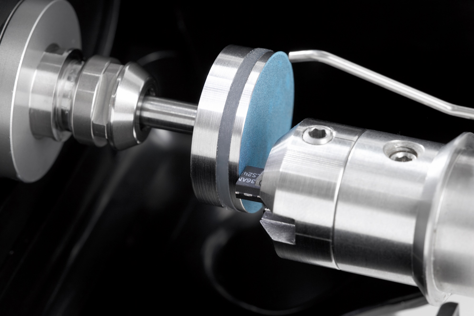
Sample Preparation for EM: A Practical Guide to Coating and Freeze-Fracturing 16 Apr 4:00 PM (2 days ago)

From coatings done in a low-vacuum sputter coating machine at room-temperature to those done in high-vacuum and even at cryogenic temperatures, Leica coating solutions cover a large range of needs. Instruments are designed to improve and optimize your sample preparation workflows from basic coatings to the most advanced freeze fracture applications.


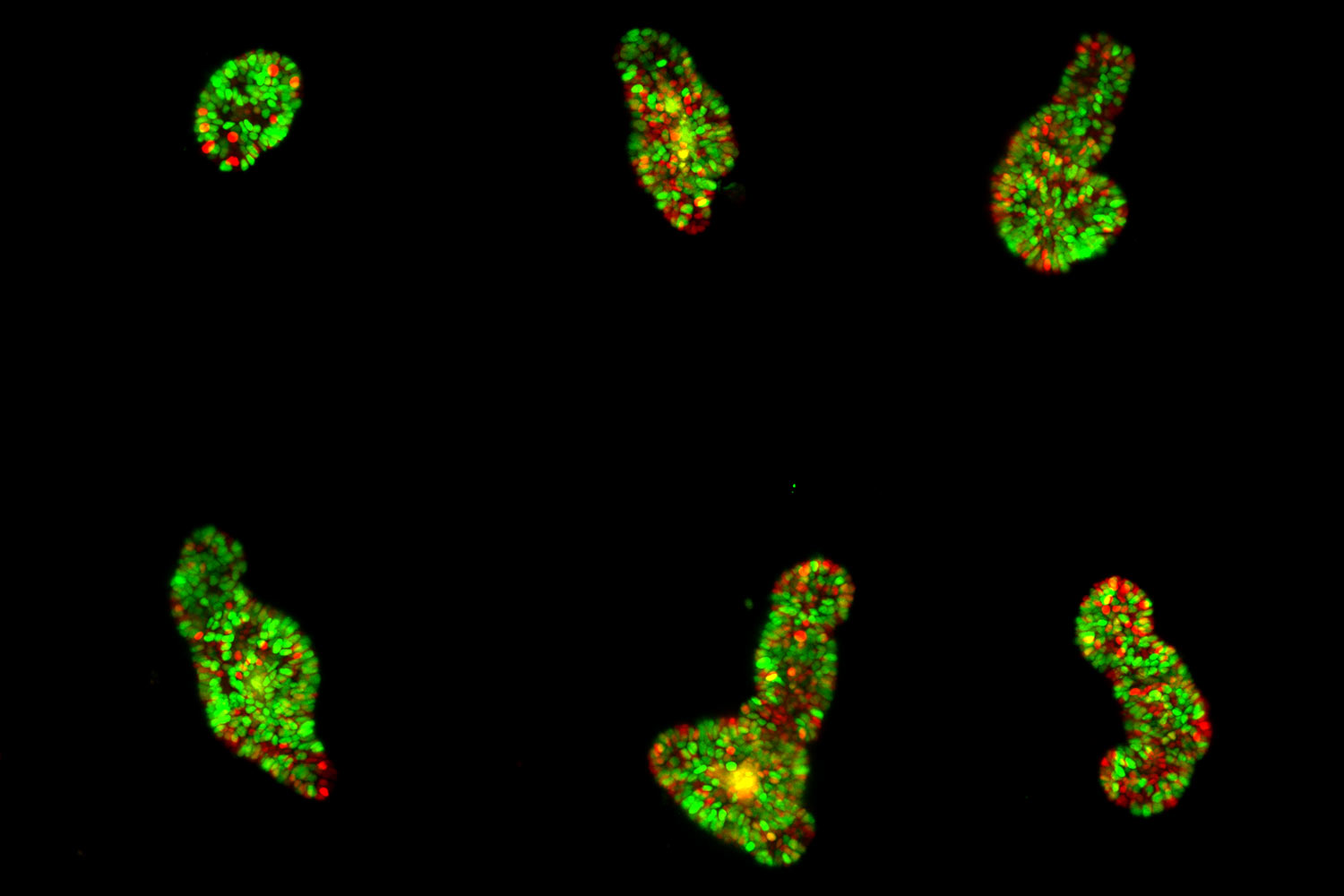
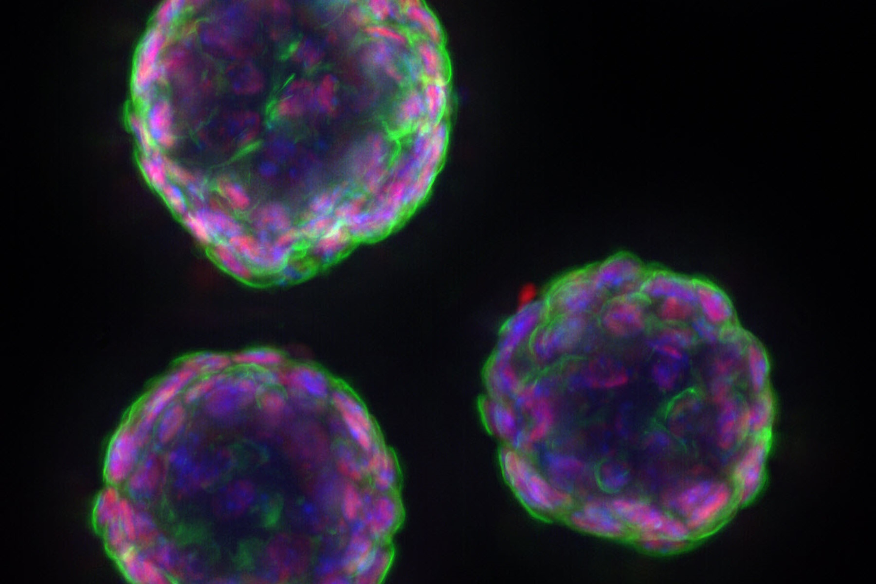
Unlocking the Secrets of Organoid Models in Biomedical Research 13 Apr 4:00 PM (5 days ago)
Get ready to delve deeper into the world of organoids and 3D models, which are essential tools for advancing our understanding of human health. Navigating these complex structures and obtaining clear images for analysis can be challenging. In this event, researchers from The University of Oxford and University College London will join us to show how the THUNDER Imager Cell Spinning Disk can provide more convincing, high-quality data for deeper insights into a variety of models.
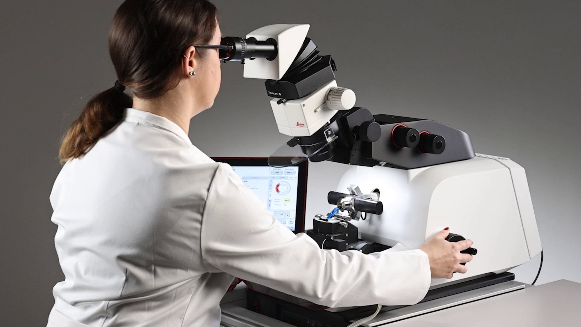
How to Save Time and Samples by Automated Ultramicrotomy 8 Apr 4:00 PM (10 days ago)
This article describes how 3D micro-CT data of a resin-embedded electron microscopy sample can be used to trim the specimen down to a defined target plane prior to sectioning. The interactive and automated approach described using Leica Microsystems’ UC Enuity saves time, reduces sample loss and training time for unexperienced users.
Essential Guide to Ultramicrotomy 3 Apr 4:00 PM (15 days ago)
When studying samples, to visualize their fine structure with nanometer scale resolution, most often electron microscopy is used. There are 2 types: scanning electron microscopy (SEM) which images the sample surface or transmission electron microscopy (TEM) which requires a very thin, electron-transparent sample. Thus, to image the fine structure inside a sample using electron microscopy, the solution is to make very thin sections of it.
Designing the Future with Novel and Scalable Stem Cell Culture 10 Mar 4:00 PM (last month)
![]()
Visionary biotech start-up Uncommon Bio is tackling one of the world’s biggest health challenges: food sustainability. In this webinar, Stem Cell Scientist Samuel East shows how they make stem cell culture media for cellular agriculture safe and economically viable. See how they achieved a 1000x reduction in media costs and developed animal free, food safe iPSC culture media.
Explore Alzheimer's Spatial Proteome with Big Data 2 Mar 3:00 PM (last month)
Alzheimer's disease, a genetic and sporadic neurodegenerative condition, leads to cognitive decline in mid to late life, marked by β-amyloid plaques and tau tangles. With limited treatment options, new investigative strategies are crucial. The Cell DIVE multiplexed imaging solution allows examination of Alzheimer's brain tissue, potentially uncovering new research avenues. Here, we showcase the Cell DIVE image viewer, enabling users to access the full Alzheimer's multiplexed dataset directly in their browser.
Automotive Part Verification and Development according to Specifications 19 Feb 3:00 PM (last month)

Automotive part verification during the development and production of parts and components by suppliers or manufacturers is important for ensuring that specifications are met. Specifications are critical for maintaining performance standards and safe operation of vehicles. However, there is always demand for efficient and cost-effective R&D and production while meeting ever-stricter quality standards. This article shows how investigation and documentation of auto parts and components to verify specifications can be done easily and quickly with digital microscopy.
Mica: A Game-changer for Collaborative Research at Imperial College London 9 Feb 3:00 PM (2 months ago)
This interview highlights the transformative impact of Mica at Imperial College London. Scientists explain how Mica has been a game-changer, expanding research possibilities and facilitating interdisciplinary collaboration. They explain how detailed live cell imaging with Mica provides more meaningful information, keeping scientists at the forefront of research. The team foresees Mica continuing to open new research avenues, including the study of microfluidics and other advanced applications.
From Bench to Beam: A Complete Correlative Cryo Light Microscopy Workflow 5 Feb 3:00 PM (2 months ago)

In the webinar entitled "A Multimodal Vitreous Crusade, a Cryo Correlative Workflow from Bench to Beam" a team of experts discusses the exciting world of correlative workflows for structural biology that empower researchers to study fine details of biological structures. Watch and explore the latest developments, instruments, and techniques in cryo workflows for correlative light electron microscopy (cryo-CLEM).
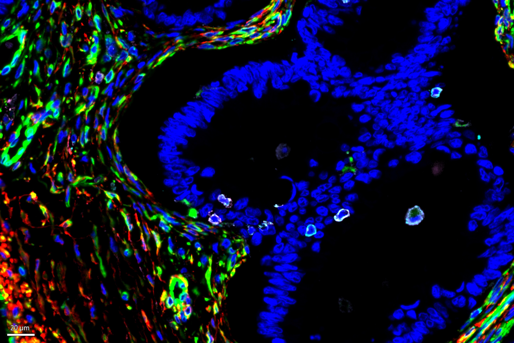
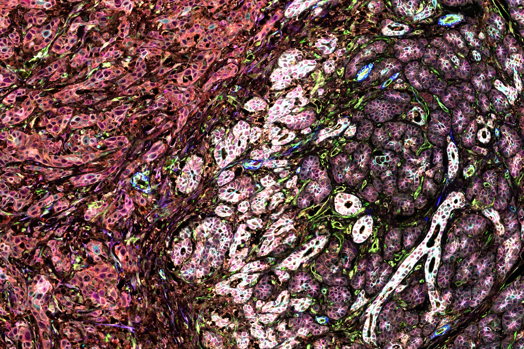
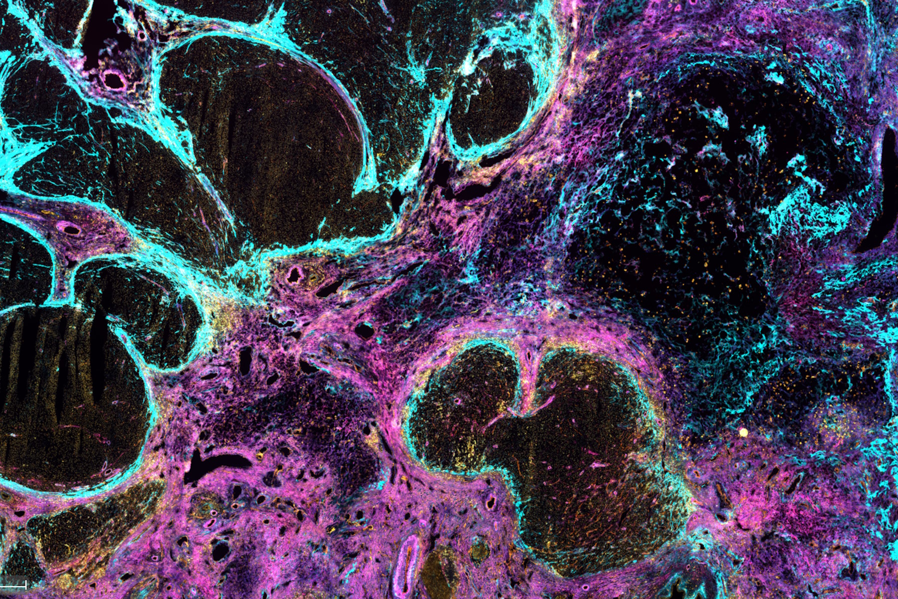
Guide to Microscopy in Cancer Research 3 Feb 3:00 PM (2 months ago)

Cancer is a complex and heterogeneous disease caused by cells deficient in growth regulation. Genetic and epigenetic changes in one or a group of cells disrupt normal function and result in autonomous, uncontrolled cell growth and proliferation.
Depth of Field in Microscope Images 3 Feb 3:00 PM (2 months ago)
For microscopy imaging, depth of field is an important parameter when needing sharp images of sample areas with structures having significant changes in depth. In practice, depth of field is determined by the correlation between numerical aperture, resolution, and magnification. For the best possible visualization of samples, modern microscopes can be adjusted to produce an optimum balance between depth of field and resolution. In theory, these parameters are inversely correlated.
Uncover the Hidden Complexity of Colon Cancer with Big Data 30 Jan 3:00 PM (2 months ago)
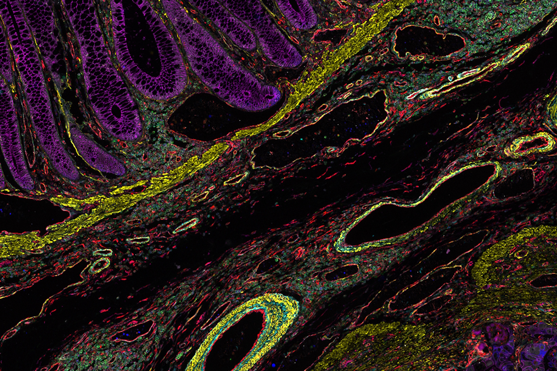
Colorectal cancer poses a significant health burden. While surgery is effective initially, some patients develop recurrent secondary disease with poor prognosis, necessitating advanced therapies like immunotherapies. Spatial biology approaches, such as multiplexed imaging with Cell DIVE, can provide crucial insights for developing novel treatments. Access the full Cell DIVE dataset in your browser to explore these findings further through the Minerva image viewer.
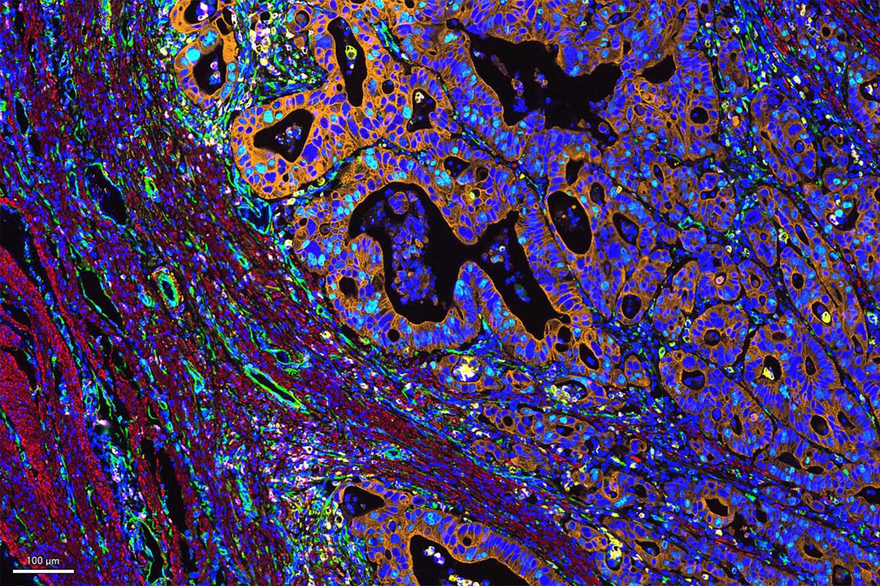
Dive into Pancreatic Cancer Research with Big Data 30 Jan 3:00 PM (2 months ago)
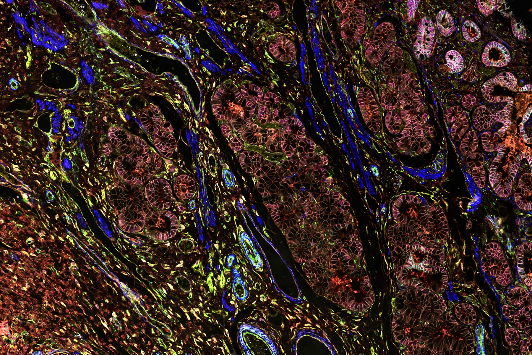
Pancreatic cancer, with a mortality rate near 40%, is challenging to treat due to its proximity to major organs. This story explores the complex biology of pancreatic ductal adenocarcinoma (PDAC), examining molecular and spatial determinants of tumor aggression in metabolism, apoptosis, and immunity. Access the full Cell DIVE dataset in your browser to delve deeper into these findings.
Introduction to 21 CFR Part 11 for Electronic Records of Cell Culture 30 Jan 12:55 AM (2 months ago)
This article provides an introduction to the recommendations of 21 CFR Part 11 from the FDA, specifically focusing on the audit trail and user management in the context of cell-culture laboratories. It is intended for professionals in the biotechnology and pharmaceutical industries who are responsible for ensuring agreement with 21 CFR part 11 for electronic records and electronic signatures. A digital microscope approach, e.g. with Mateo FL, offers the advantage of more consistent and efficient electronic documentation of cell-culture results compared to a paper-based method.
A Guide to Phase Contrast 22 Jan 3:00 PM (2 months ago)
A phase contrast light microscope offers a way to view the structures of many types of biological specimens in greater contrast without the need of stains.
A Guide to Super-Resolution 22 Jan 3:00 PM (2 months ago)
Find out more about Leica super-resolution microscopy solutions and how they can empower you to visualize in fine detail subcellular structures and dynamics.
Overcoming Challenges with Microscopy when Imaging Moving Zebrafish Larvae 21 Jan 3:00 PM (2 months ago)
Zebrafish is a valuable model organism with many beneficial traits. However, imaging a full organism poses challenges as it is not stationary. Here, this case study shows how zebrafish larvae can be imaged during stationary periods and easily relocated after movement. The seamlessly integrated widefield and confocal capability of Mica is leveraged to capture fast events, like the heartbeat, with virtually no out-of-focus background noise which is inherent to standard widefield systems.
Selecting the Right Dissecting Microscope 21 Jan 3:00 PM (2 months ago)

You can spend many hours looking through the eyepieces of a dissecting microscope whenever dissections must be done. Leica Microsystems lets you select from a diverse array of microscopes and comprehensive range of dissecting microscope parts and accessories so that you can find the microscope solution that best fits your needs.
A Guide to Neuroscience Research 21 Jan 3:00 PM (2 months ago)

Are you working towards a better understanding of neurodegenerative diseases or studying the function of the nervous system? See how you can make breakthroughs with imaging solutions from Leica Microsystems.
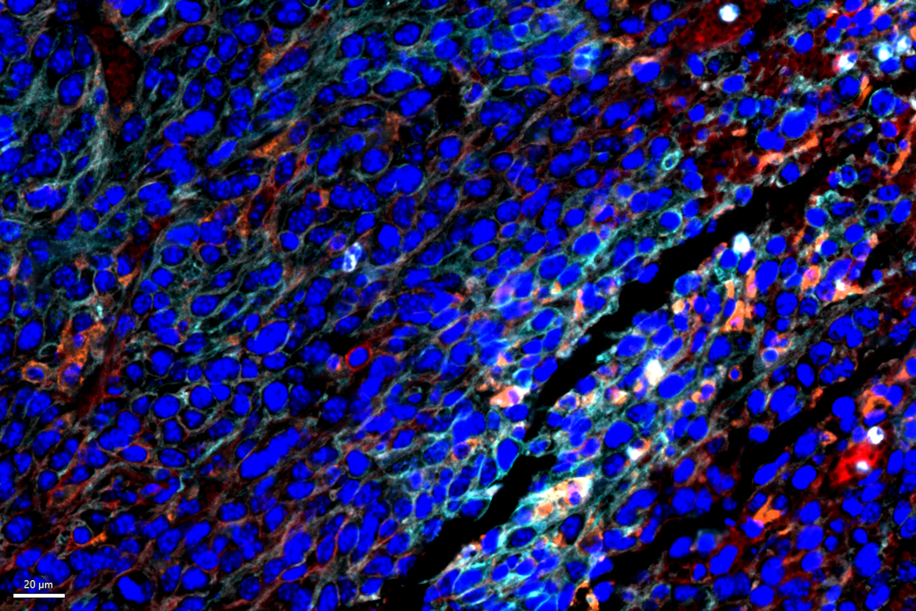
Mapping Tumor Immune Landscape with AI-Powered Spatial Proteomics 20 Jan 3:00 PM (2 months ago)
Spatial mapping of untreated tumors provides an overview of the tumor immune architecture, useful for understanding therapeutic responses. Immunocompetent murine models are essential for identifying immune-dependent events in tumor development and progression. Characterizing these models, with intact immune systems and interacting cellular components, requires a multiplexed approach. We show an AI-powered spatial proteomics approach to study the tumor-immune interactions in murine cancer tissue.
A Practical Guide to Virology Research 19 Jan 3:00 PM (2 months ago)
Leica solutions for imaging and sample preparation help you with the investigation of viral entry and fusion, genome integration, viral replication, assembly, and virus budding.
A Guide to Zebrafish Research 16 Jan 3:00 PM (3 months ago)
For the best result during screening, sorting, manipulation, and imaging you need to see details and structures to make the right decisions for your next steps in research.
Known for outstanding optics and superb resolution, stereo microscopes and transmitted light bases from Leica are the preferred choice of researchers worldwide.
A Guide to Cryo-Electron Tomography 16 Jan 3:00 PM (3 months ago)
Cryo-electron tomography (CryoET) is used to resolve biomolecules within their cellular environment down to an unprecedented resolution below one nanometer.
A Guide to OCT 16 Jan 3:00 PM (3 months ago)

Leica Optical Coherence Tomography (OCT) systems support ophthalmologists, ophthalmic surgeons, and researchers with easy-to-use, high-quality imaging technology.
Aneurysm Clipping: Assessing Perforators in Real-Time with AR Fluorescence 13 Jan 3:00 PM (3 months ago)
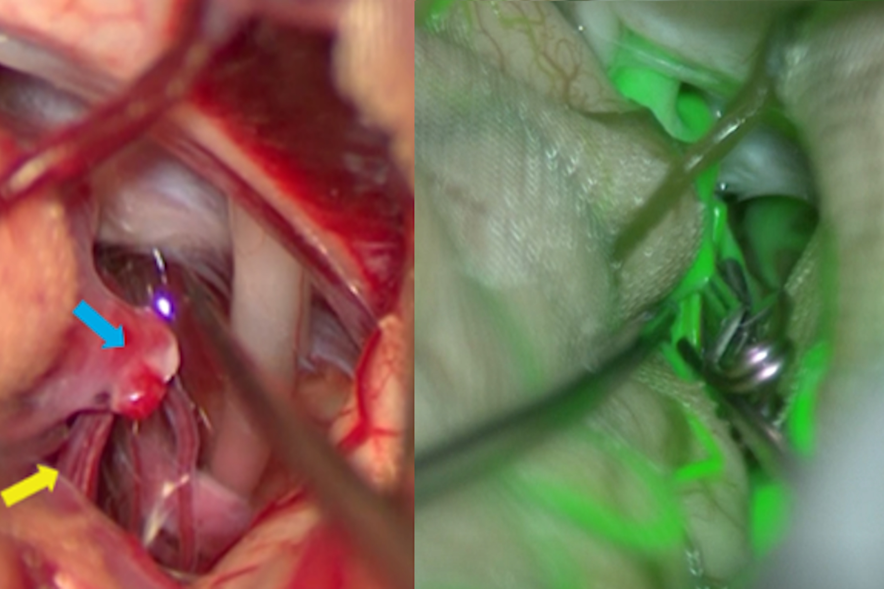
This article covers two aneurysm clipping cases highlighting the clinical benefits of GLOW800 Augmented Reality Fluorescence application in neurosurgery, based on insights from Prof. Tohru Mizutani, Chief Professor of the Department of Neurosurgery at Showa University Hospital (Japan). It shows how a neurosurgeon can visualize in 3D the real-time blood flow correlated with the anatomy in natural colour and with depth perception during aneurysm clipping and other complex neurosurgical techniques.
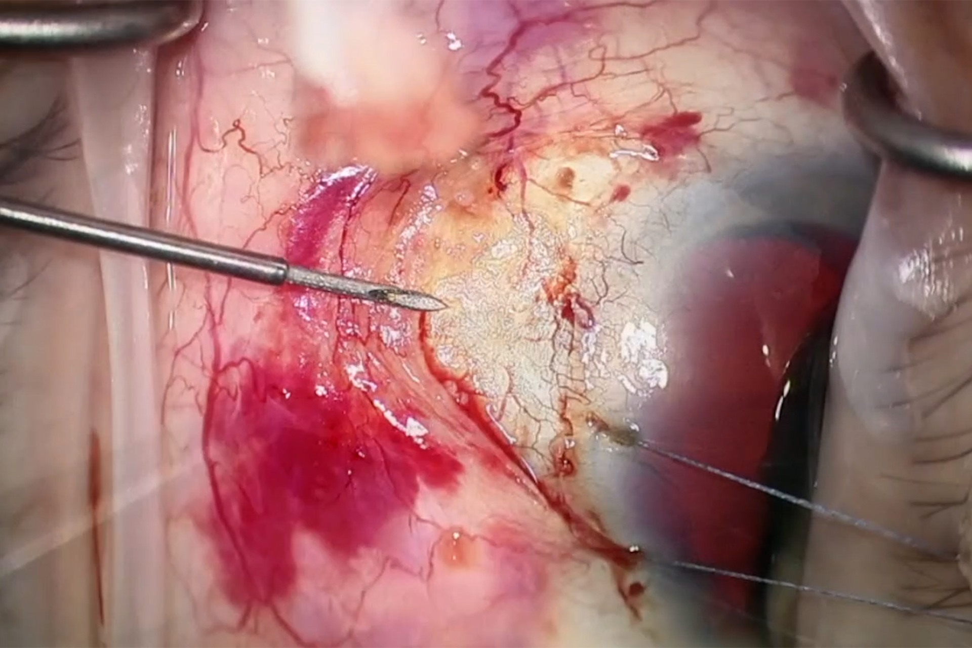
How does Real-Time OCT Imaging Impact Precision in Corneal Surgery? 12 Jan 3:00 PM (3 months ago)
Corneal surgery is a highly specialized field. It requires great surgical precision to overcome challenges such as visualizing clearly the full anterior chamber, performing Descemet membrane peeling and endothelium procedures, measuring how deep to cut the corneal stroma, and quantifying the incision depth as well as avoiding damage to the donor tissue. Watch how Prof. Fontana uses real-time intraoperative OCT imaging during cornea transplantation procedures to enhance his surgical precision.
How to Achieve Brain Tissue Resection with GLOW400 AR 12 Jan 3:00 PM (3 months ago)

Intraoperative MRI is one form of real-time intraoperative visualization, but if more in-depth visualization to identify a tumor during surgery is wanted, intraoperative fluorescence diagnostics is available. In this article, we interviewed Professor Akihide Kondo about his clinical experience of glioma surgery with the GLOW400 Augmented Reality fluorescence application for brain tumor surgery from Leica Microsystems.
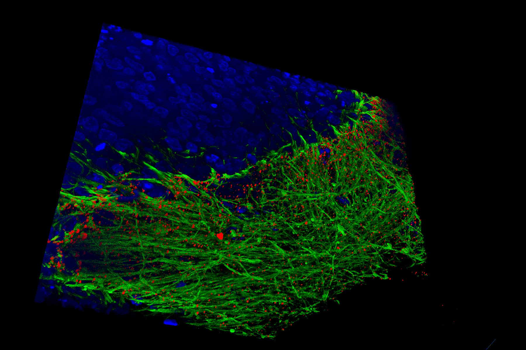
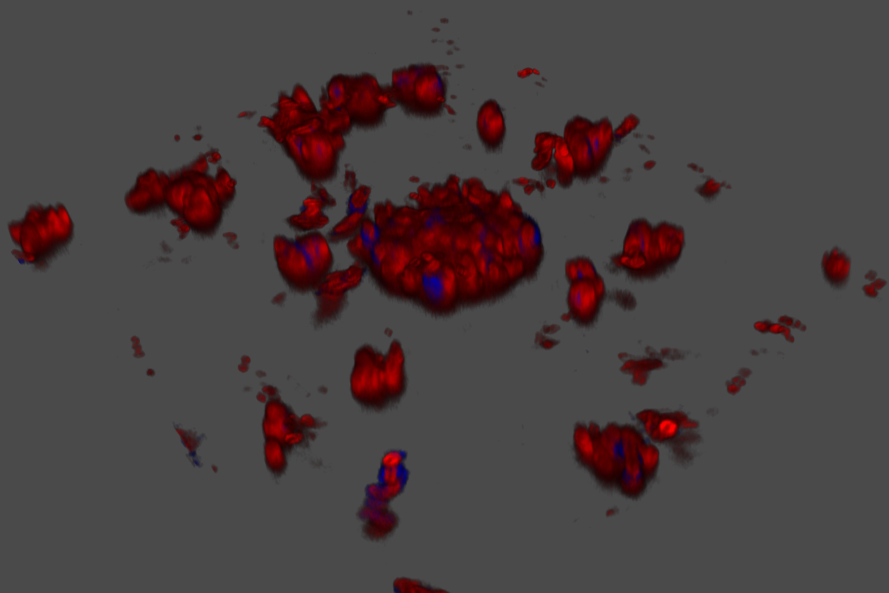
How to Automatically Obtain Fluorescent Cells of Interest in a Block-face 12 Dec 2024 3:00 PM (4 months ago)
Block-face created by automatic trimming under fluorescence.
Mammalian cells of interest, stained with CellTrackerTM Green are visualized within the block-face using the UC Enuity equipped with the stereo microscope M205 FA. In the background a carbon finder grid in black is visible.
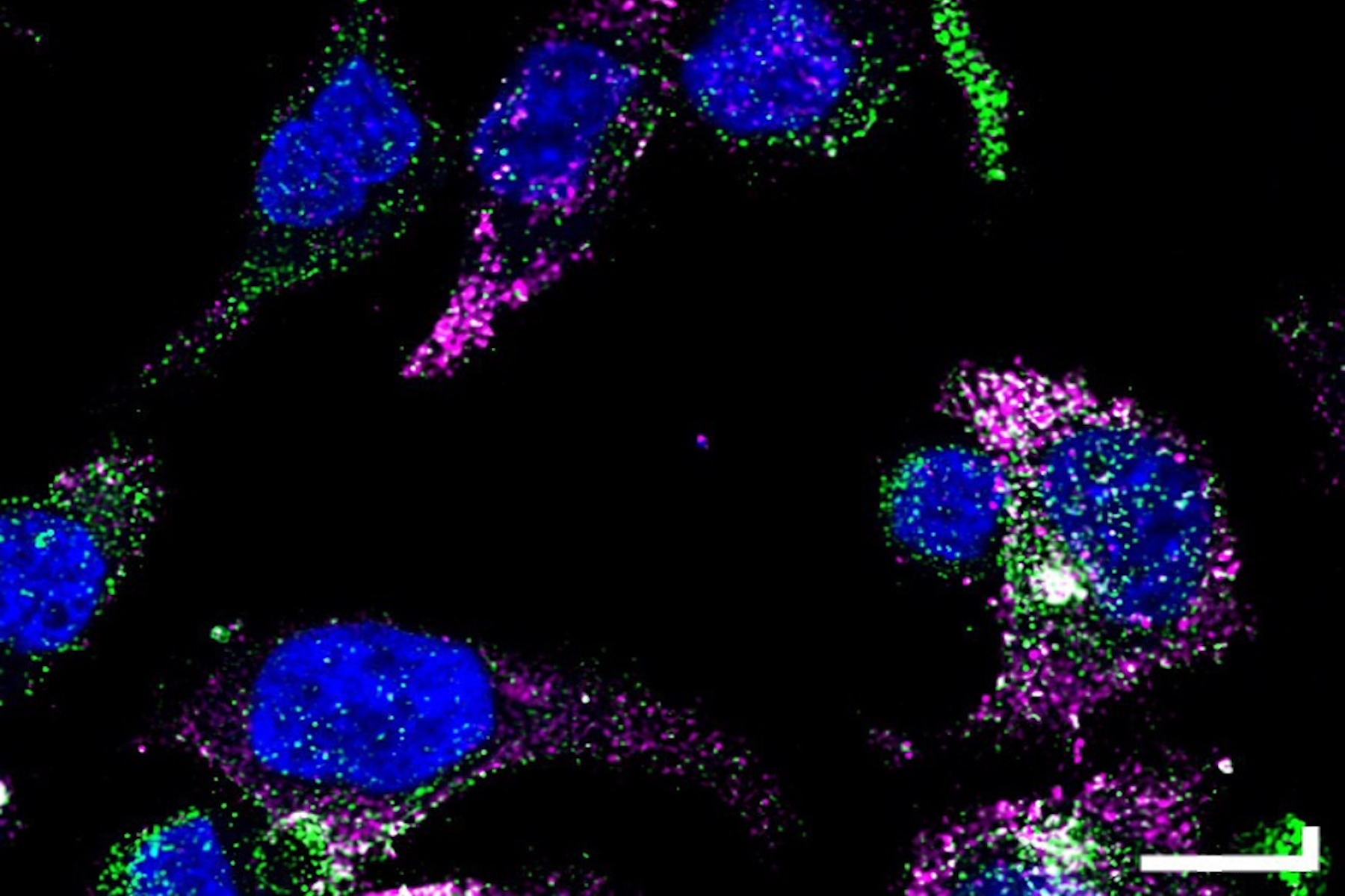
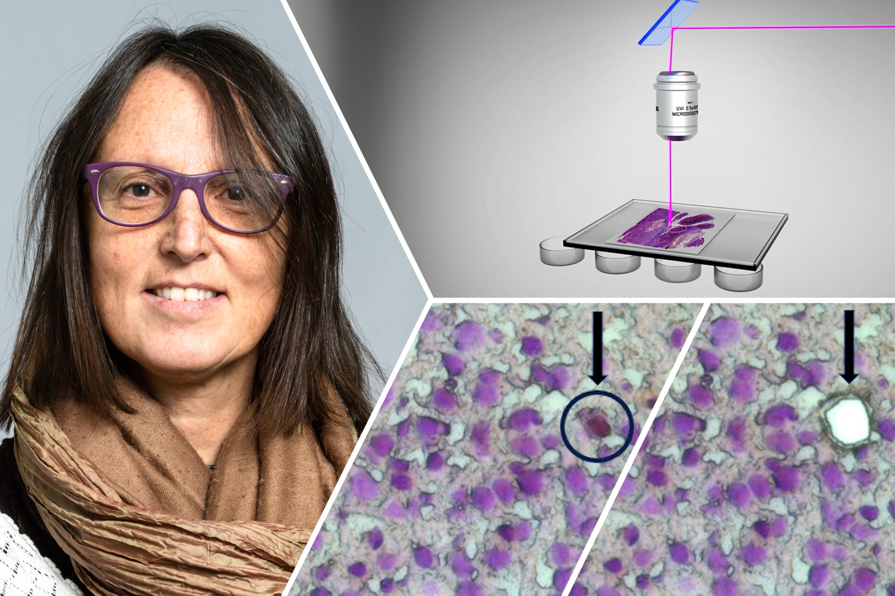
Deep Visual Proteomics Provides Precise Spatial Proteomic Information 1 Dec 2024 3:00 PM (4 months ago)
Despite the availability of imaging methods and mass spectroscopy for spatial proteomics, a key challenge that remains is correlating images with single-cell resolution to protein-abundance measurements. Deep Visual Proteomics (DVP), a recent method, combines artificial-intelligence-driven image analysis of cellular phenotypes with automated single-cell or single-nucleus laser microdissection and ultra-high-sensitivity mass spectrometry. DVP links protein abundance to complex cellular or subcellular phenotypes while preserving spatial context.
What is Empty Magnification and How can Users Avoid it 20 Nov 2024 3:00 PM (4 months ago)

The phenomenon of “empty magnification”, which can occur while using an optical, light, or digital microscope, and how it can be avoided is explained in this article. The performance of an optical microscope is highly dependent on the numerical aperture, wavelength of light, and resolution, not just the magnification. However, there is often a relationship between numerical aperture and magnification. From the numerical aperture and light wavelength, the range over which the magnification is useful can be determined. Going beyond that range results in empty magnification.
How to Study Gene Regulatory Networks in Embryonic Development 20 Nov 2024 12:11 AM (5 months ago)

Join Dr. Andrea Boni by attending this on-demand webinar to explore how light-sheet microscopy revolutionizes developmental biology. This advanced imaging technique allows for high-speed, volumetric live imaging of 3D samples with minimal phototoxicity. Learn through user examples how light-sheet microscopy is enhancing our understanding of intestinal and brain organoid development and dive into the technology behind the Viventis Deep microscope from Leica Microsystems and its application in long-term imaging.
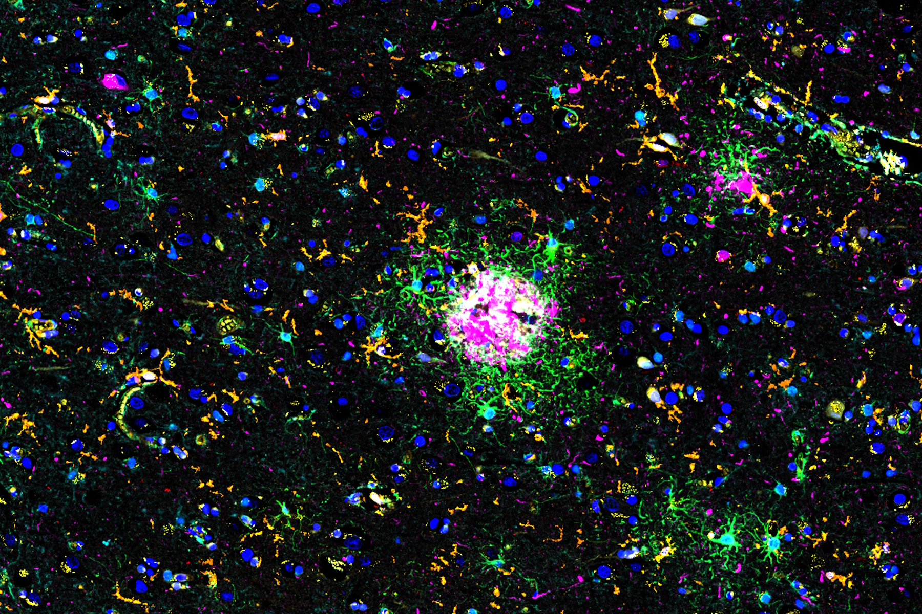
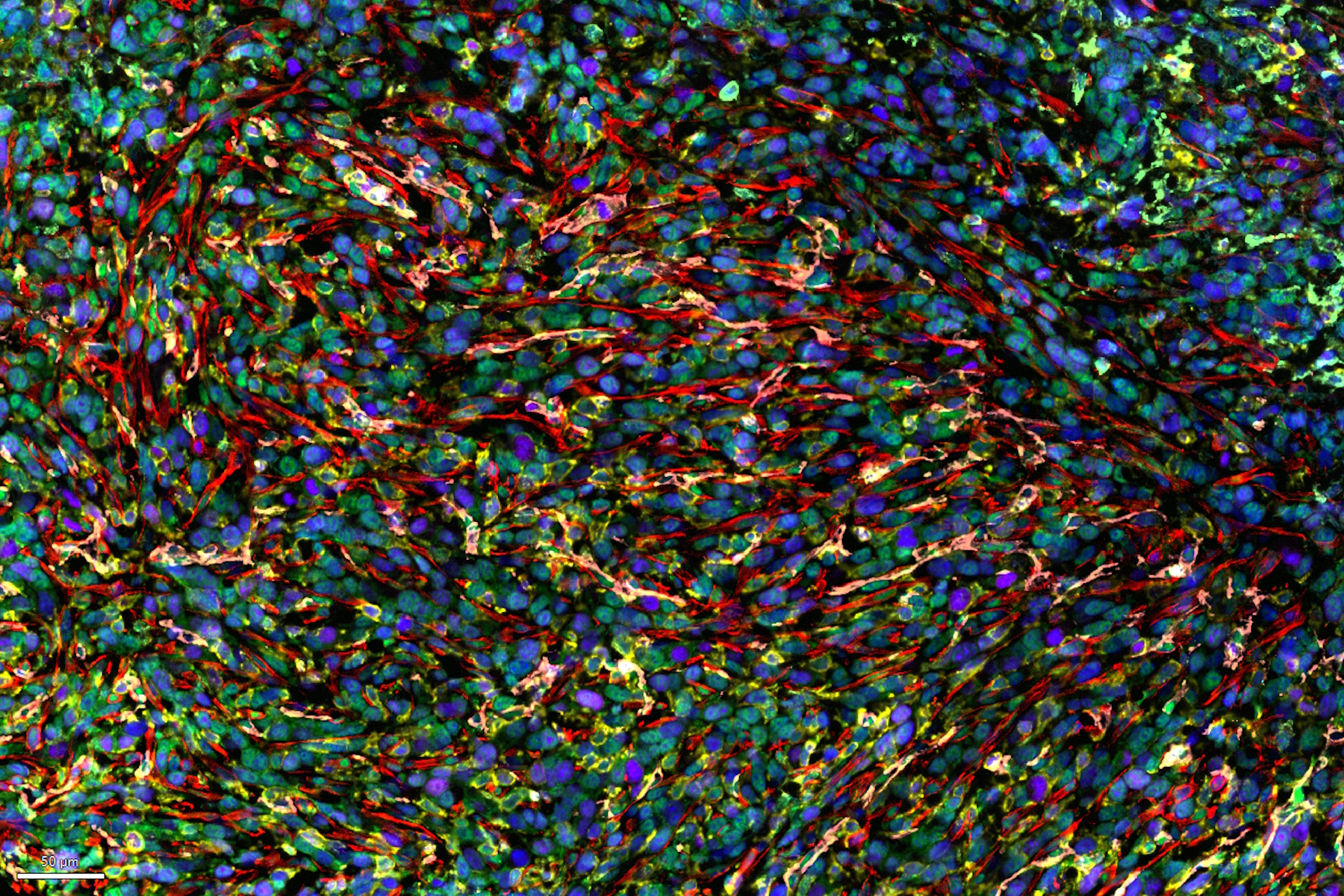
Spatial Analysis of Neuroimmune Interactions in Alzheimer’s Disease 10 Nov 2024 3:00 PM (5 months ago)
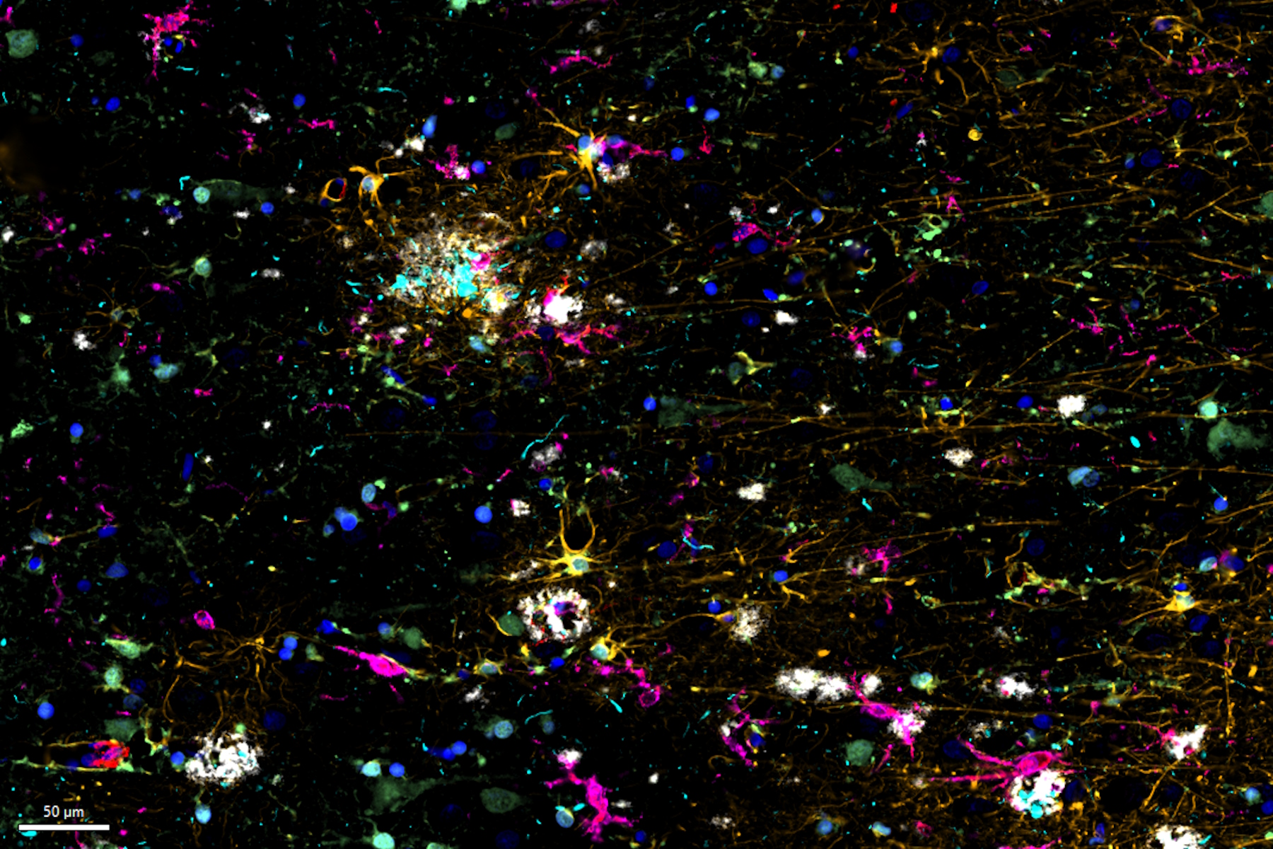
Alzheimer’s disease (AD) is a complex neurodegenerative disorder characterized by neurofibrillary tangles, β-amyloid plaques, and neuroinflammation. These dysfunctions trigger or are exacerbated by local immune responses. Therefore, understanding neuroimmune interactions with spatial context is crucial for elucidating AD pathogenesis. Here we utilize multiplexed imaging with Cell DIVE and AI-assisted spatial analysis using Aivia to study immune cells surrounding pathological hallmarks in AD.
The Polarization Microscopy Principle 30 Oct 2024 11:08 PM (5 months ago)

Polarization microscopy is routinely used in the material and earth sciences to identify materials and minerals on the basis of their characteristic refractive properties and colors. In biology, polarization microscopes are commonly used for identification of birefringent structures, like crystals, or for imaging of cellulose in the walls of plant cells and starch grains. This article gives an overview of the basic principles of polarization microscopy.
A Guide to Spatial Biology 10 Oct 2024 12:54 AM (6 months ago)

What is spatial biology, and how can researchers leverage its tools to meet the growing demands of biological questions in the post-omics era? This article provides a brief overview of spatial biology and its technologies, as well as key research questions in this dynamic field.
An Introduction to Laser Microdissection 8 Oct 2024 11:39 PM (6 months ago)

The heterogeneity of histological and biological specimens often requires isolation of specific single cells or cell groups from surrounding tissue before molecular biology analysis can be carried out. Laser microdissection (LMD) is a highly selective process for preparing samples of DNA, RNA, protein, or other biomaterials for analysis. It is a microscope-controlled manipulation technique for the precise separation of samples, cells, and tissue using a focused laser beam. This article explains the basic principles of LMD.
Cutting-Edge Imaging Techniques for GPCR Signaling 2 Oct 2024 12:54 AM (6 months ago)
With this webinar on-demand enhance your pharmacological research with our webinar on GPCR signaling and explore cutting-edge imaging techniques that aim to understand how GPCR signaling translates into cellular and physiological responses. Discover leading research that's expanding what we know about these critical pathways to find new avenues for drug discovery.
Revealing Neuronal Migration’s Molecular Secrets 29 Sep 2024 10:06 PM (6 months ago)

Different approaches can be used to investigate neuronal migration to their niche in the developing brain. In this webinar, experts from The University of Oxford present the microscopy tools and assays they use to elucidate the molecular mechanisms of neuronal migration to functional layers of the cortex during neurodevelopment. Understanding these processes will lead to a better understanding of healthy brain development and potentially better treatments for neurodevelopmental disorders.
Exploring Microbial Worlds: Spatial Interactions in 3D Food Matrices 24 Sep 2024 2:24 AM (6 months ago)
The Micalis Institute is a joint research unit in collaboration with INRAE, AgroParisTech, and Université Paris-Saclay. Its mission is to develop innovative research in the field of food microbiology for health. In this video series, researchers from the Micalis Institute explain how Mica is used in various microscopy experiments to investigate the interactions between food, foodborne microorganisms, and the host. Using Mica,enables rapid, high-resolution 3D imaging of food matrices and microbial interactions, they can better understand the emerging properties of these spatially organized communities.
Molecular Biology Analysis facilitated with Laser Microdissection (LMD) 18 Sep 2024 5:00 AM (7 months ago)

Extracting biomolecules, proteins, nucleic acids, lipids, and chromosomes, as well as extracting and manipulating cells and tissues with laser microdissection (LMD) enables insights to be gained into the function of genes and proteins. It is used for a range of applications in neurobiology, immunology, developmental, cell biology, and forensics, e.g., research on cancer and diseases, genetic modifications, molecular pathology and biology, and biochemistry. LMD is also useful for studying protein functions, molecular mechanisms, and their interaction in transduction pathways.
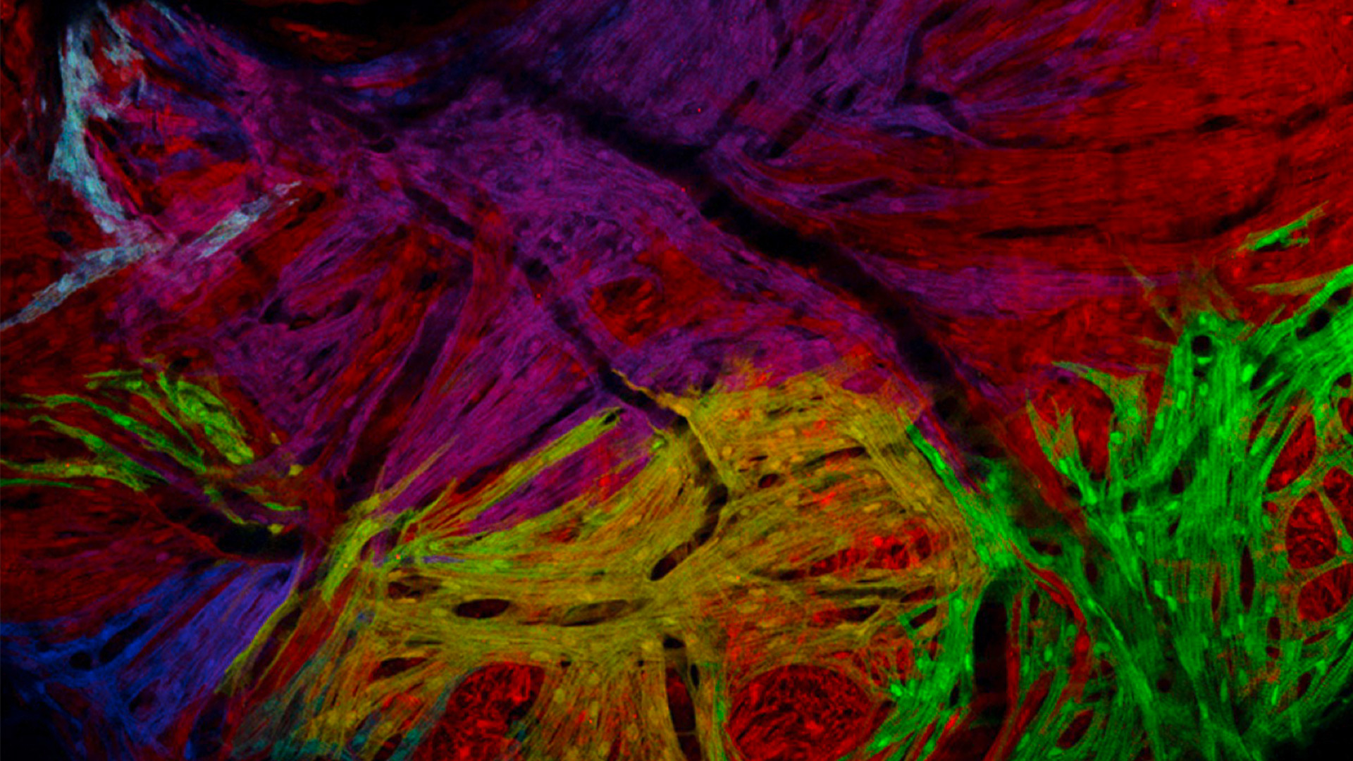
Probing Human Alzheimer's Cortical Section using Spatial Multiplexing 16 Sep 2024 11:42 PM (7 months ago)
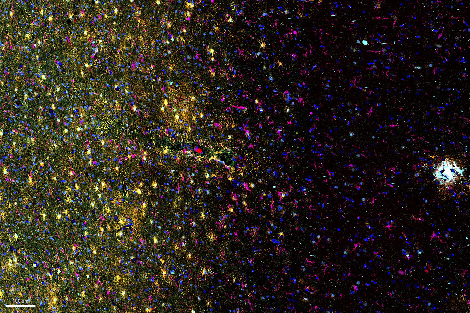
Alzheimer’s disease (AD) is the most common neurodegenerative disease and is characterized by the progressive decline of cognitive function. Spatial profiling of AD brain may reveal cellular relationships, facilitating a better understanding of disease etiology. This study captures a global overview of the AD cortical tissue composition and emphasizes the streamlined workflow of Cell DIVE imaging, from data acquisition to AI-based analysis using Aivia software, resulting in quicker insights.
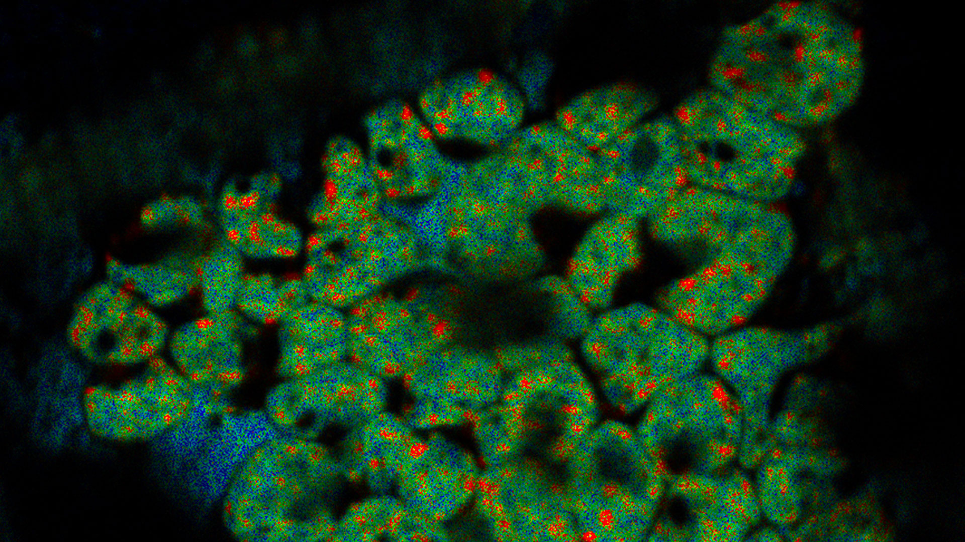
Spatial Metabolomics: Exploring Tumor Complexity and Therapeutic Insights 3 Sep 2024 12:14 AM (7 months ago)
In cancer research, it is vital to understand the interaction between tumor cells and their microenvironment, as the tumor microenvironment influences tumor progression significantly. Spatial Metabolomics is a novel method developed by researchers to investigate this complexity. By uncovering spatial variations within the tumor microenvironment, this method provides valuable insights into its diverse components and their organization. These insights not only impact clinical outcomes but also inform therapeutic responses, paving the way for personalized treatment strategies.
Advancing Uterine Regenerative Therapies with Endometrial Organoids 2 Sep 2024 4:00 PM (7 months ago)
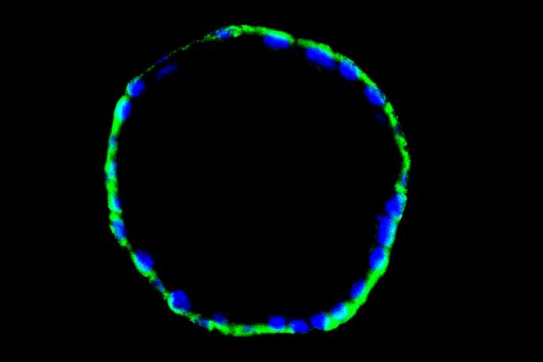
Prof. Kang's group investigates important factors that determine the uterine microenvironment in which embryo insertion and pregnancy are successfully maintained. They are working to develop new therapeutic strategies that regenerate endometrial function for patients suffering from endometrial disease like Asherman’s syndrome. Her group transplants 3D endometrial organoids into mouse models to identify the cellular and molecular mechanisms that underlie the remarkable regenerative capacity of endometrium. Find out from this interview what kind of research her group is doing and how Mica has been helpful.
Lipidomics Analysis of Sparse Cells based on Laser Microdissection 27 Aug 2024 12:49 AM (7 months ago)
Delve into cellular intricacies with high-coverage targeted lipidomics analysis of sparse cells. This advanced method, integrating Laser Microdissection (LMD) and Liquid Chromatography-Mass Spectrometry/Mass Spectrometry (LC-MS/MS), reveals metabolic changes at the single-cell level, shedding light on diseases like diabetes and obesity. By employing Laser Microdissection (LMD) for contamination-free samples and the SCIEX 7500 system for increased sensitivity, this method successfully detects 285 lipids across seven classes in isolated single cells. This precision extends to other cell section samples marking a significant leap in precision metabolomics research. This article explores the nuanced landscape of cellular metabolism through this approach.
Rapidly Visualizing Magnetic Domains in Steel with Kerr Microscopy 14 Aug 2024 1:07 AM (8 months ago)

The rotation of polarized light after interaction with magnetic domains in a material, known as the Kerr effect, enables the investigation of magnetized samples with Kerr microscopy. It allows rapid visualization of magnetic domains at the material’s surface. For efficient R&D and QC of magnetic materials, e.g., steel alloys, used in electrical and electronic devices, Kerr microscopy can play an important role. More details about how Kerr microscopy can be used to image magnetic domains in the grains of steel alloys is described in this article.
How Efficient is your 3D Organoid Imaging and Analysis Workflow? 5 Aug 2024 11:51 PM (8 months ago)

Organoid models have transformed life science research but optimizing image analysis protocols remains a key challenge. This webinar explores a streamlined workflow for organoid research, starting with real-time 3D cell culture checks. This is followed by high-speed, high-resolution 3D imaging to generate crisp images and cleaner data for accurate AI-based segmentation and quantification of parameters such as growth rate, cell migration and 3D cellular interactions – enabling deeper insights.
Improve Your Ultramicrotomy Workflow with Automated Sectioning 31 Jul 2024 11:51 PM (8 months ago)

Discover advanced digital ultramicrotomy tools for fast and accurate automated sectioning. Learn about autoalignment, and efficient sample trimming leveraging 3D µCT data. See application examples that advance EM sample preparation workflows. Watch now to enhance your lab's precision and efficiency.
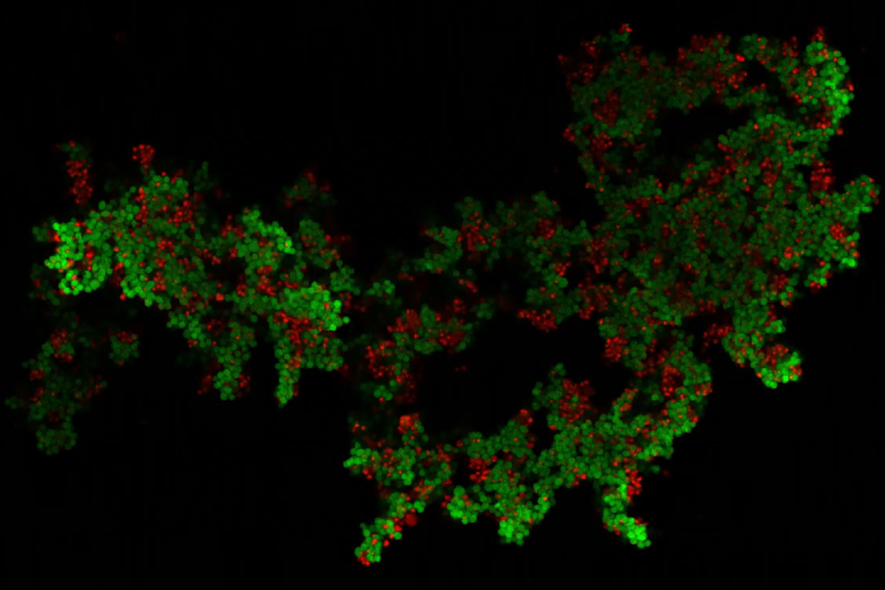
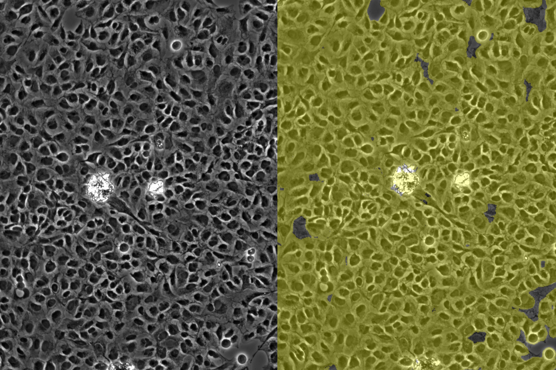
Leveraging AI for Efficient Analysis of Cell Transfection 26 Jul 2024 6:00 AM (8 months ago)
![]()
This article explores the pivotal role of artificial intelligence (AI) in optimizing transfection efficiency measurements within the context of 2D cell culture studies. Precise and reliable transfection efficiency measurements for 2D cell culture are key for understanding cellular mechanisms. A high transfection efficiency of the targeted proteins is crucial for experiments including live-cell imaging and protein purification. Manual estimation is inconsistent and unreliable. With the power of AI, efficient and reliable transfection studies can be achieved.
Precision and Efficiency with AI-Enhanced Cell Counting 26 Jul 2024 6:00 AM (8 months ago)
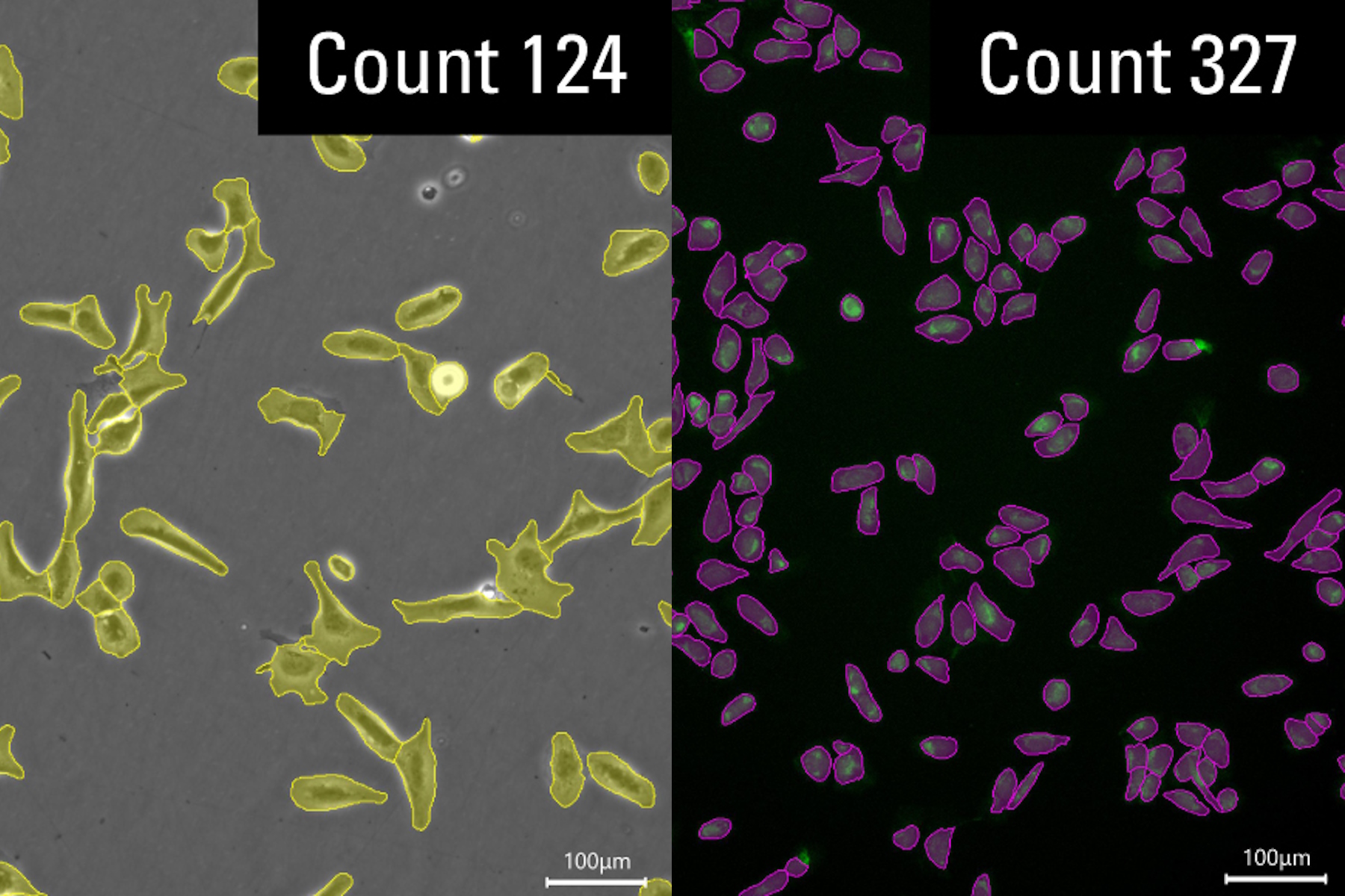
This article describes the use of artificial intelligence (AI) for precise and efficient cell counting. Accurate cell counting is important for research with 2D cell cultures, e.g., cellular dynamics, drug discovery, and disease modelling. Precise cell counting is critical for determining cell viability, proliferation rates, and the effects of experimental conditions. These factors are essential for reliable and robust results. How an AI-based approach can significantly enhance the accuracy and speed of cell counting, leading to a significant impact on cellular research, is described.
AI Confluency Analysis for Enhanced Precision in 2D Cell Culture 26 Jul 2024 6:00 AM (8 months ago)
This article explains how efficient, precise confluency assessment of 2D cell culture can be done with artificial intelligence (AI). Assessing confluency, the percentage of surface area covered, accurately is crucial for reliable cell research. Traditional methods use visual inspection or simplistic algorithms, making precision challenging, especially with complex cell lines used for drug discovery, tissue engineering, and regenerative medicine. Methods exploiting AI with automated image analysis and deep-learning algorithms offer better precision and can enhance experimental results.
Empowering Spatial Biology with Open Multiplexing and Cell DIVE 17 Jul 2024 2:22 AM (9 months ago)

Spatial biology and multiplexed imaging workflows have become important in immuno-oncology research. Many researchers struggle with study efficiency, even with effective tools and protocols. Here, we describe how researchers used the adaptability of open multiplexing to combine IBEX imaging with Cell DIVE, creating a technique called Cell DIVE-IBEX. It allows these researchers to adapt existing techniques and reagents and gain the scalability of Cell DIVE for their immuno-oncology study.
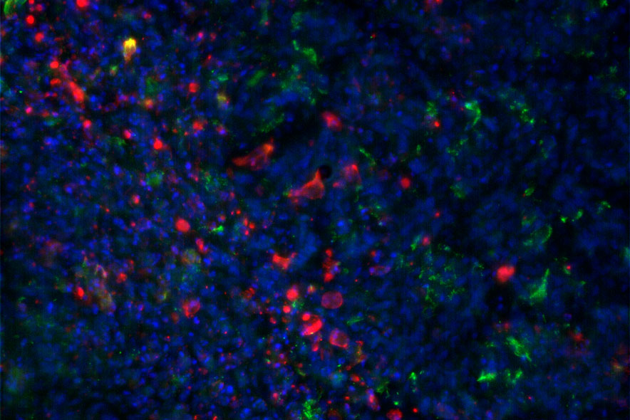
Neuron Isolation in Spatial Context with Laser Microdissection (LMD) 24 Jun 2024 3:11 AM (9 months ago)

After Alzheimer’s disease, Parkinson’s is the second most common progressive neurodegenerative disease. Before the first symptoms manifest, up to 70% of dopamine-releasing neurons in the mid-brain have already died. This article describes how modern laser microdissection (LMD) methods are used to help solve the Parkinson’s disease puzzle. The research involves neuron isolation and analysis in a spatial context. The cells are from substantia-nigra tissue specimens taken from postmortem Parkinson’s patients in order to gain molecular insight into the disease.
How did Laser Microdissection enable Pioneering Neuroscience Research? 24 Jun 2024 12:04 AM (9 months ago)
Dr. Marta Paterlini, a Senior Scientist at the Karolinska Institute, shares her experience of using laser microdissection (LMD) in groundbreaking research into adult human neurogenesis and offers personal insights into its potential future applications in spatial proteomics and precision medicine.
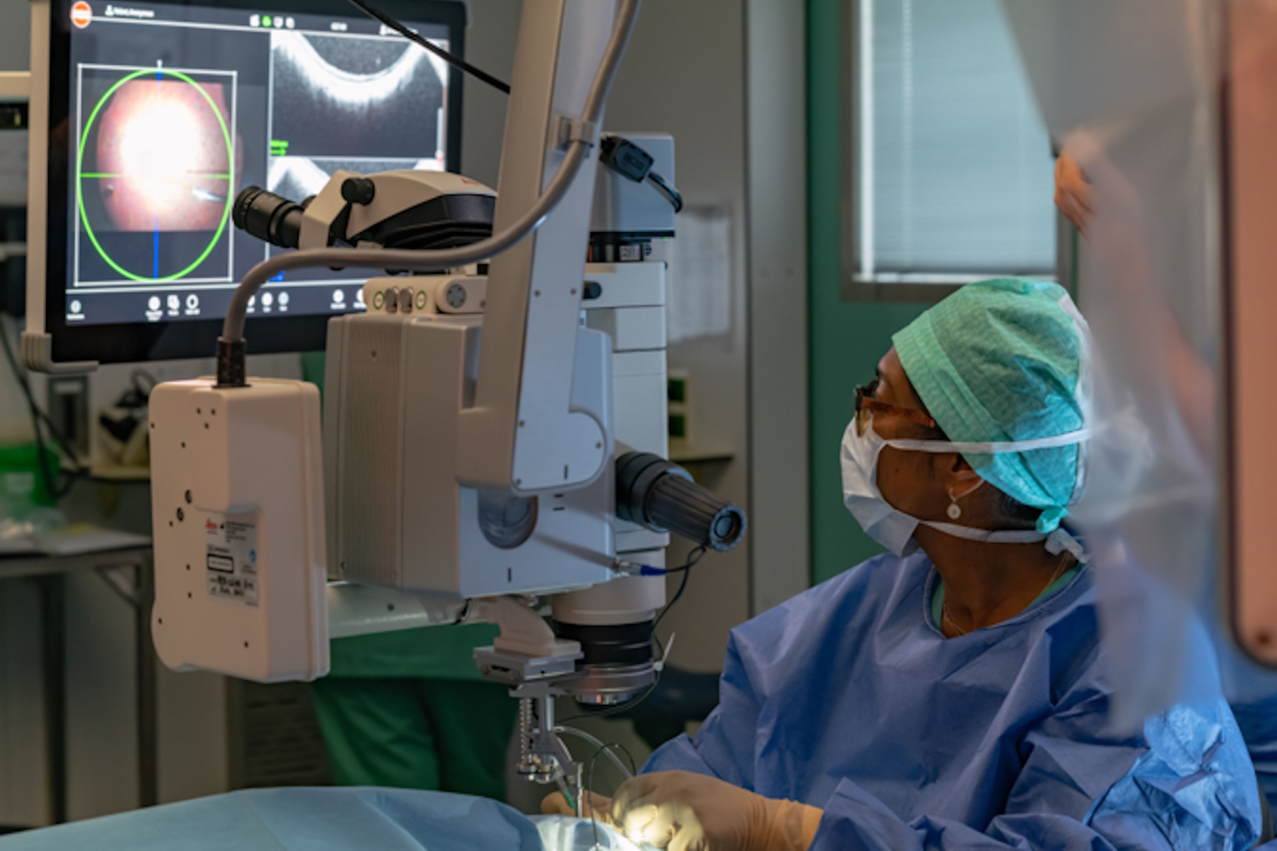
Buying an Ophthalmic Microscope? Gain Peers Insights from Dr. Dhami 12 Jun 2024 11:26 PM (10 months ago)
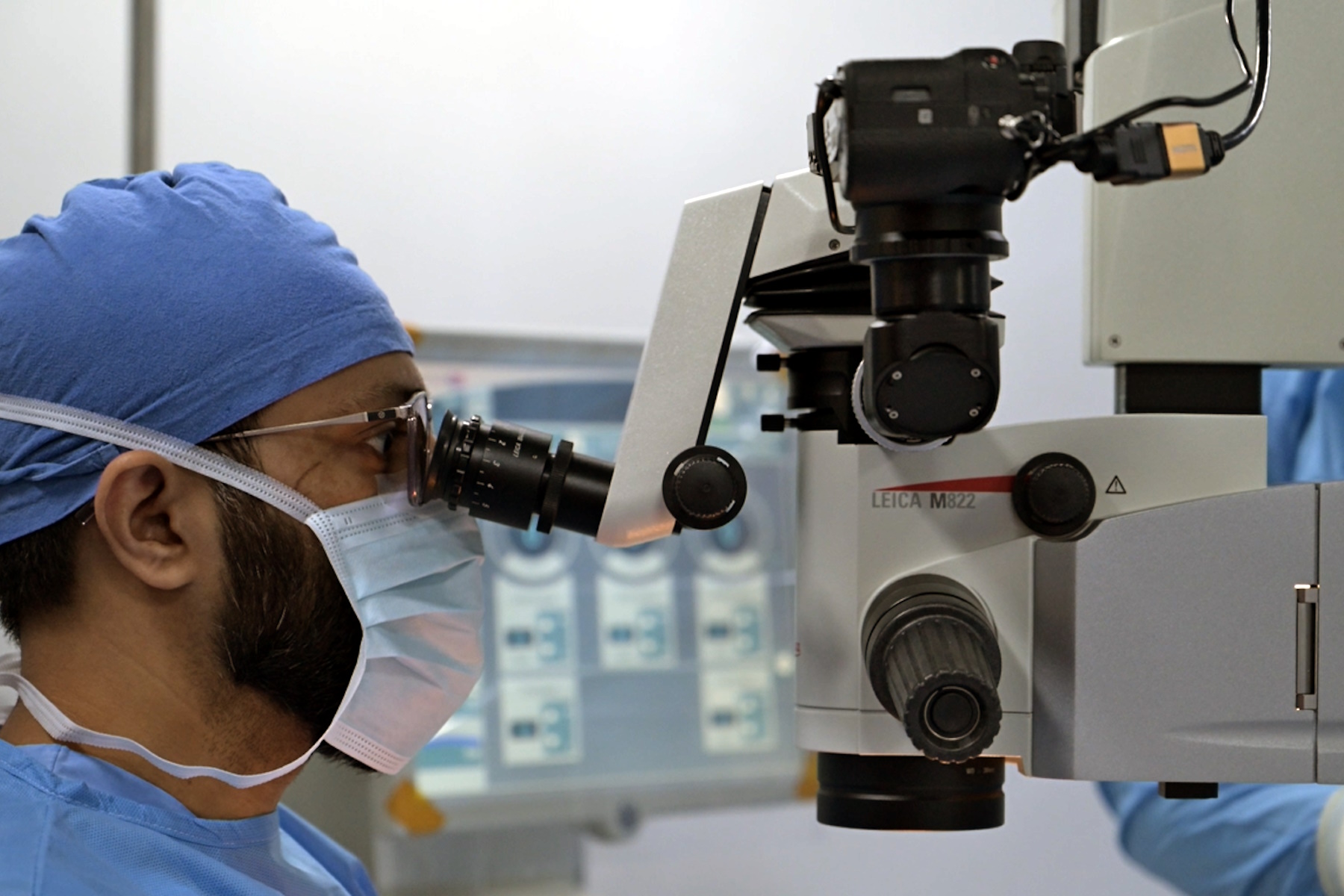
In this article, learn how Dr. Abhinav Dhami, an ophthalmic surgery consultant in North India, enhances his surgical precision using the M822 ophthalmic microscope from Leica Microsystems and the key features that convinced him to buy this ophthalmic microscope for his practice.
6-Inch Wafer Inspection Microscope for Reliably Observing Small Height Differences 12 Jun 2024 4:11 AM (10 months ago)

A 6-inch wafer inspection microscope with automated and reproducible DIC (differential interference contrast) imaging, no matter the skill level of users, is described in this article. Manufacturing of integrated-circuit (IC) chips and semiconductor components requires wafer inspection to verify no defects are present which affect performance. The inspection is often done with optical microscopy for QC, failure analysis, and R&D. To visualize efficiently small height differences between structures on wafers, DIC can be used.
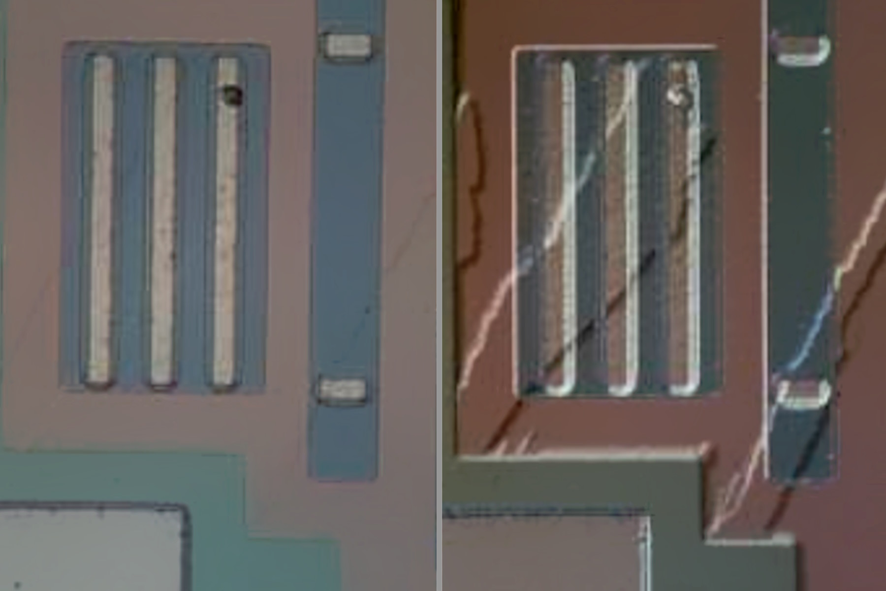
Visualizing Photoresist Residue and Organic Contamination on Wafers 11 Jun 2024 11:59 PM (10 months ago)

As the scale of integrated circuits (ICs) on semiconductors passes below 10 nm, efficient detection of organic contamination, like photoresist residue, and defects during wafer inspection is becoming more crucial. Optical microscopy is still the common inspection method, but for organic contamination brightfield and other types of illumination can have limitations. How fluorescence microscopy is used to efficiently detect photoresist residues and other organic contamination on wafers during QC, failure analysis, and R&D for the semiconductor industry is discussed in this article.
Augmented Reality: Transforming Neurosurgical Procedures 11 Jun 2024 3:54 AM (10 months ago)

In this ebook, you will explore the exciting advances that Augmented Reality (AR) brings to the field of neurosurgery. This comprehensive guide, including explanatory videos, addresses key questions and provides detailed explanations, shedding light on the future of surgical practices.
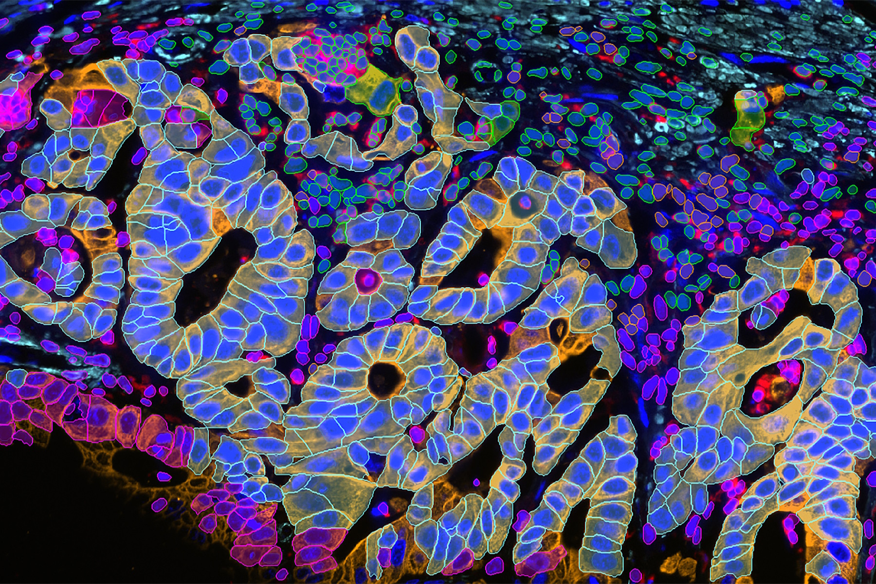
AI-Powered Multiplexed Image Analysis to Explore Colon Adenocarcinoma 3 Jun 2024 12:14 AM (10 months ago)
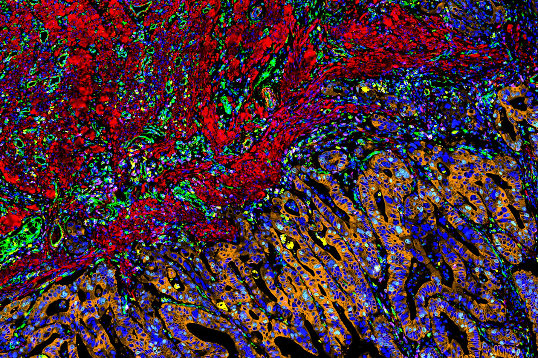
In this application note, we demonstrate a spatial biology workflow via an AI-powered multiplexed image analysis-based exploration of the tumor immune microenvironment in colon adenocarcinoma.
Laser Microdissection Protocols for Tissue and Cell Isolation - Download free eBook 21 May 2024 9:28 PM (11 months ago)

In this Bio-protocol Selections, we present a collection of open-access, detailed methods papers using LCM to purify and isolate tissues and cells from plants, mouse embryos, cancer cells, neurons, and more.
How do Cells Talk to Each Other During Neurodevelopment? 21 May 2024 4:19 AM (11 months ago)

Professor Silvia Capello presents her group’s research on cellular crosstalk in neurodevelopmental disorders, using models such as cerebral organoids and assembloids.
Tympanoplasty Surgery: Optimal Approaches and Tools 21 May 2024 1:00 AM (11 months ago)

Discover tympanoplasty surgery case studies illustrating the standard approaches: post-auricular, endaural and transcanal. Gain insights from Dr. Flanagan on selecting the adequate surgical approach and right visualization tools for successful tympanic membrane perforation repair.
Workflow Solutions for Sample Preparation Methods for Material Science 18 May 2024 4:00 PM (11 months ago)

This brochure presents and explains appropriate workflow solutions for the most frequently required sample preparation methods for material science samples.
Dual-View LightSheet Microscope for Large Multicellular Systems 7 May 2024 1:38 AM (11 months ago)
Visualizing the dynamics of complex multicellular systems is a fundamental goal in biology. To address the challenges of live imaging over large spatiotemporal scales, Franziska Moos et. al. present an open-top multisample dual-view lightsheet microscope in a paper published in Nature Methods. The authors find that the Viventis Deep microscope offers significant advances in imaging large samples with single-cell resolution.
A Meta-cancer Analysis of the Tumor Spatial Microenvironment 25 Apr 2024 10:58 PM (11 months ago)
Learn how clustering analysis of Cell DIVE datasets in Aivia can be used to understand tissue-specific and pan-cancer mechanisms of cancer progression
Mapping the Landscape of Colorectal Adenocarcinoma with Imaging and AI 25 Apr 2024 10:24 PM (11 months ago)
Discover deep insights in colon adenocarcinoma and other immuno-oncology realms through the potent combination of multiplexed imaging of Cell DIVE and Aivia AI-based image analysis
Spatial Architecture of Tumor and Immune Cells in Tumor Tissues 25 Apr 2024 10:16 PM (11 months ago)
Dig deep into the spatial biology of cancer progression and mouse immune-oncology in this poster, and learn how tumor metabolism can effect immune cell function.
IBEX, Cell DIVE, and RNA-Seq: A Multi-omics Approach to Follicular Lymphoma 7 Apr 2024 11:19 PM (last year)
In a recent study by Radtke et al., a multi-omics spatial biology approach helps shed light on early relapsing lymphoma patients
Overcoming Observational Challenges in Organoid 3D Cell Culture 7 Apr 2024 10:48 PM (last year)

Learn how to overcome challenges in observing organoid growth. Read this article and discover new solutions for real-time monitoring which do not disturb the 3D structure of the organoids over time.
Burr Detection During Battery Manufacturing 3 Apr 2024 11:16 PM (last year)

See how optical microscopy can be used for burr detection on battery electrodes and determination of damage potential to achieve rapid and reliable quality control during battery manufacturing.
Battery Particle Detection During the Production Process 3 Apr 2024 3:43 AM (last year)

How battery particle detection and analysis is enhanced with optical microscopy and laser spectroscopy for rapid, reliable, and cost-effective QC during battery production is explained in this article.
Rapid Check of Live Stem Cells in Cell-Culture Inserts set in Multi-Well Plates 20 Mar 2024 2:47 AM (last year)
See how efficient imaging of live iPSC stem cells within cell-culture inserts set in a multi-well plate can be done to evaluate the cells using a THUNDER Imager. Just read this article.
Automatic Alignment of Sample and Knife for High Sectioning Quality 19 Mar 2024 3:30 AM (last year)
Automatic alignment of sample and knife on the ultramicrotome UC Enuity, enabling even untrained users to create ultrathin sections with reduced risk of losing precious sections.
High Quality Sectioning in Ultramicrotomy 19 Mar 2024 12:56 AM (last year)

Discover the significance of achieving high-quality uniform sections with ultramicrotomy for precise imaging in electron microscopy.
Super-Resolution Microscopy Image Gallery 14 Mar 2024 4:00 PM (last year)

Due to the diffraction limit of light, traditional confocal microscopy cannot resolve structures below ~240 nm. Super-resolution microscopy techniques, such as STED, PALM or STORM or some deconvolution processing methods, are used when enhanced resolution is needed to study structures and molecular events below the diffraction limit scale.
Extended Live-cell Imaging at Nanoscale Resolution 4 Mar 2024 11:48 PM (last year)

Extended live-cell imaging with TauSTED Xtend. Combined spatial and lifetime information allow super-resolution microscopy at extremely low light dose.
The Guide to STED Sample Preparation 4 Mar 2024 10:00 PM (last year)

This guide is intended to help users optimize sample preparation for stimulated emission depletion (STED) nanoscopy, specifically when using the STED microscope from Leica Microsystems. It gives an overview of fluorescent labels used for single color STED imaging and a ranking of their performance.
How to Streamline Your Histology Workflows 27 Feb 2024 9:52 PM (last year)

Streamline your histology workflows. The unique Fluosync detection method embedded into Mica enables high-res RGB color imaging in one shot.
How to Prepare Samples for Stimulated Raman Scattering (SRS) imaging 4 Feb 2024 11:59 PM (last year)

Find here guidelines for how to prepare samples for stimulated Raman scattering (SRS), acquire images, analyze data, and develop suitable workflows. SRS spectroscopic imaging is also known as SRS microscopy.
RPE65 Gene Therapy Subretinal Injection: Benefits of Intraoperative OCT 31 Jan 2024 11:18 PM (last year)
Discover how RPE65 gene therapy subretinal injection procedures in patients with Leber congenital amaurosis is supported by intraoperative Optical Coherence Tomography.
Accelerating Discovery for Multiplexed Imaging of Diverse Tissues 30 Jan 2024 9:45 PM (last year)

Explore IBEX: Open-source multiplexed imaging. Join the collaborative IBEX Imaging Community for optimized tissue processing, antibody selection, and human atlas construction.
Key Factors for Efficient Cleanliness Analysis 2 Jan 2024 10:27 PM (last year)

An overview of the key factors necessary for technical cleanliness and efficient cleanliness analysis concerning automotive and electronics manufacturing and production is provided in this article.
Technical Terms for Digital Microscope Cameras and Image Analysis 13 Dec 2023 11:39 PM (last year)

Learn more about the basic principles behind digital microscope camera technologies, how digital cameras work, and take advantage of a reference list of technical terms from this article.
Rapid Semiconductor Inspection with Microscope Contrast Methods 12 Dec 2023 10:21 PM (last year)
Semiconductor inspection during the production of patterned wafers and ICs (integrated circuits) is important for identifying and minimizing defects. To increase the efficiency of quality control in the early stages of production and to ensure reliable IC chip performance, microscopy solutions should combine different contrast methods that provide complete and accurate information about different defects. Our free guide details microscopy techniques & optimizing quality control. Get your free copy today!
Transforming Multiplexed 2D Data into Spatial Insights Guided by AI 12 Dec 2023 10:08 PM (last year)
Aivia 13 handles large 2D images and enables researchers to obtain deep insights into microenvironment surrounding their phenotypes with millions of detected objects and automatic clustering up to 30 markers.
Dislocated Cataract Angle Closure Aided by Intraoperative OCT 6 Dec 2023 9:01 PM (last year)
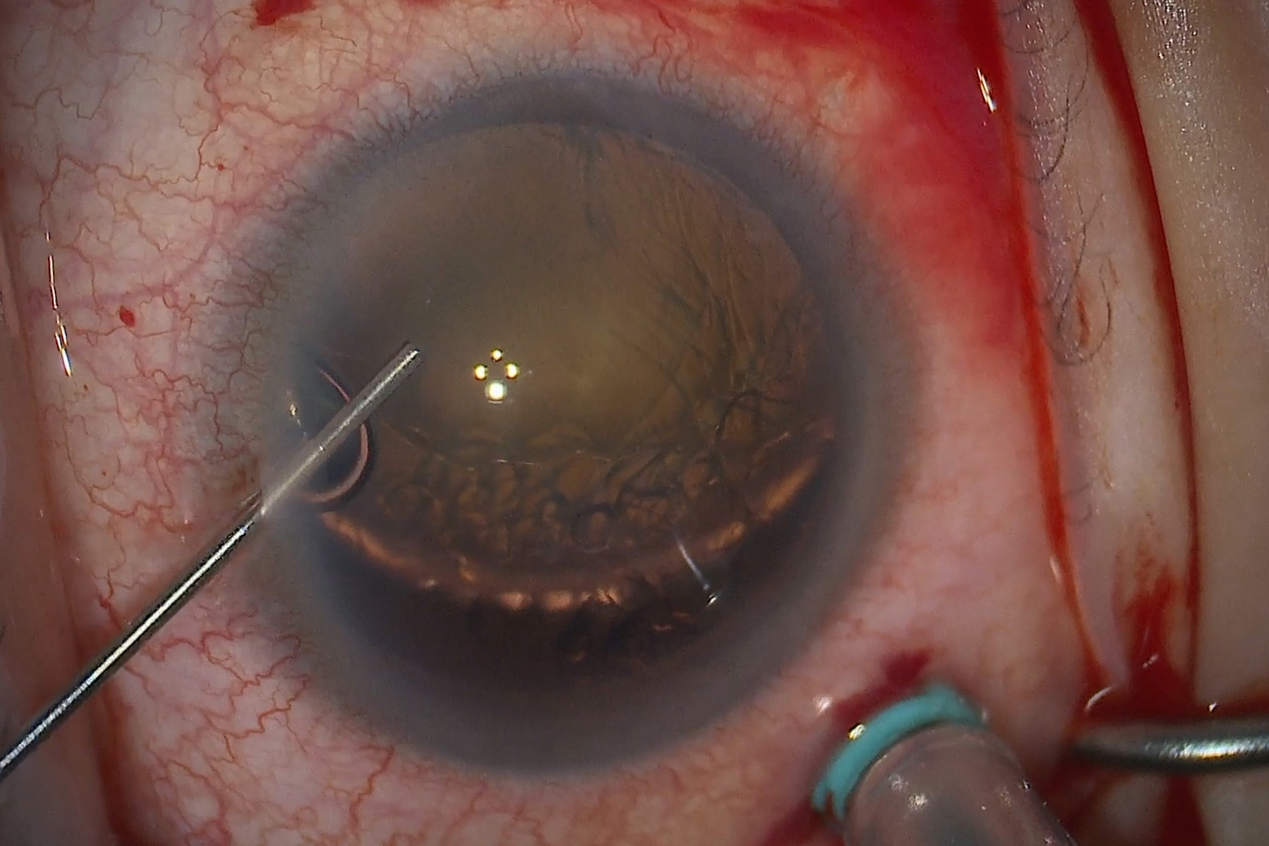
Learn how a dislocated cataract was treated with angle closure assisted by intraoperative OCT to achieve long-term good results without future lens dislocation.
Notable AI-based Solutions for Phenotypic Drug Screening 5 Dec 2023 11:55 PM (last year)

Learn about notable optical microscope solutions for phenotypic drug screening using 3D-cell culture, both planning and execution, from this free, on-demand webinar.
Understanding Tumor Heterogeneity with Protein Marker Imaging 3 Dec 2023 11:46 PM (last year)
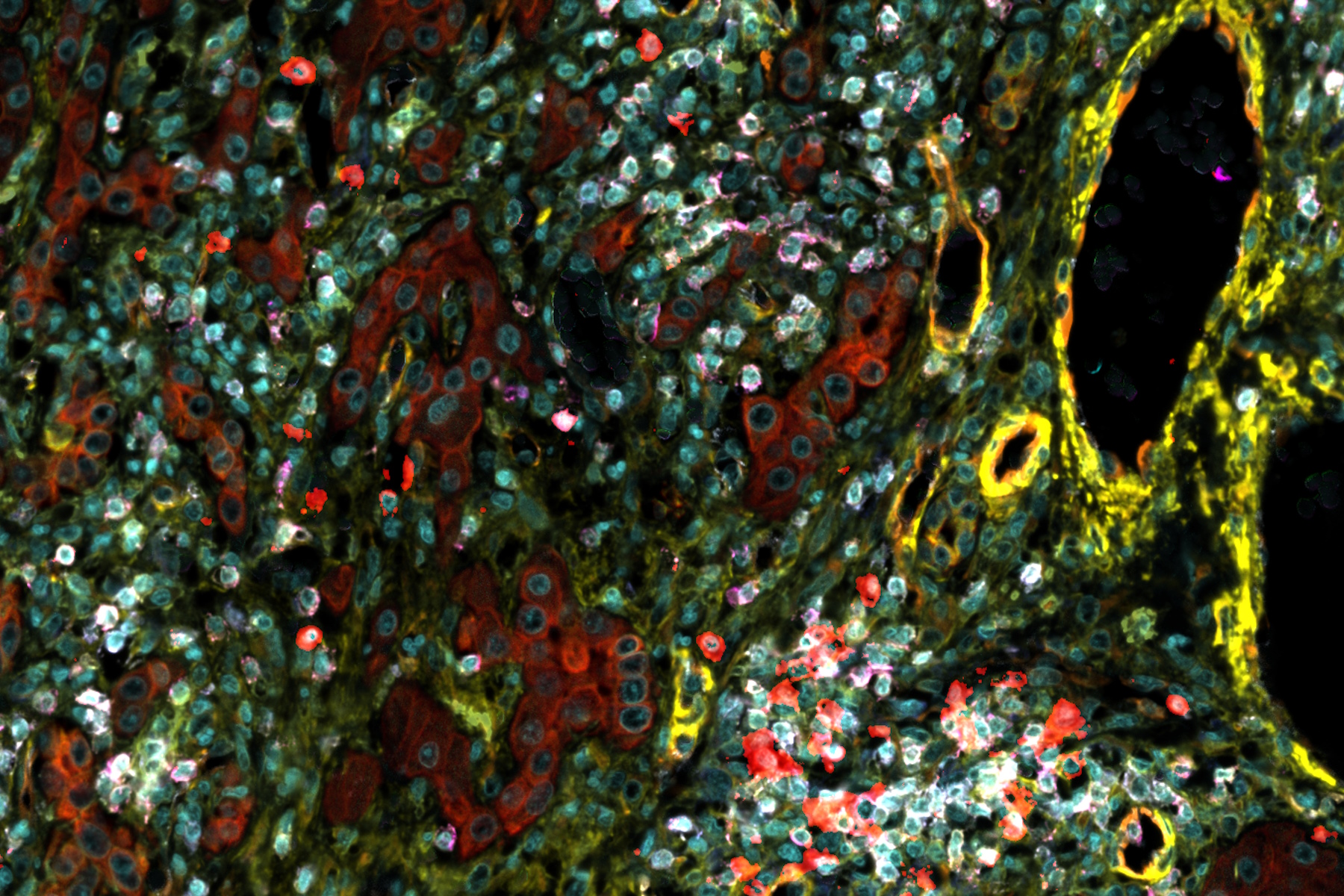
Explore tumor heterogeneity and immune cell dynamics. See how quantitative imaging analysis reveals spatial relationships and molecular insights crucial for advancing cancer research and therapeutics.
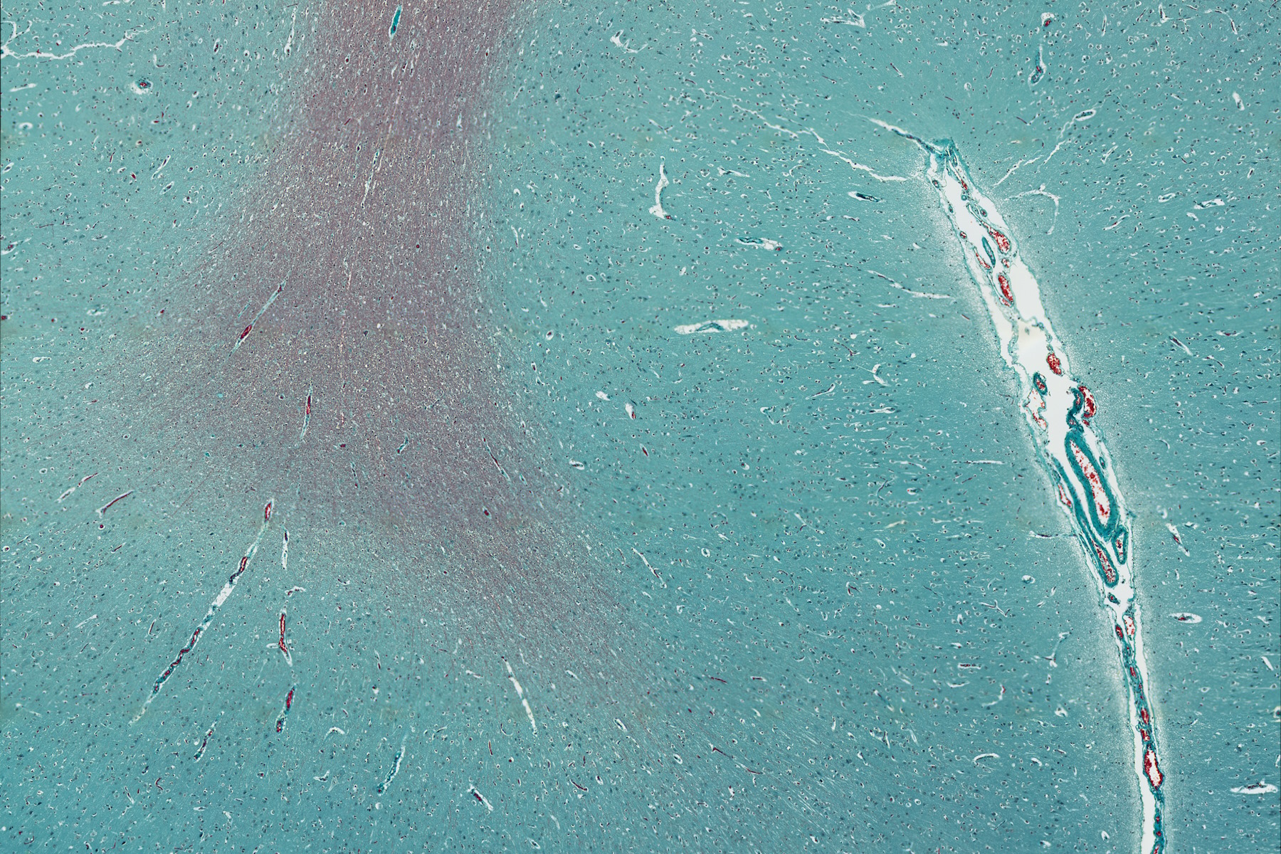
The Shape of the Brain: Spatial Biology of Alzheimer’s Disease 28 Nov 2023 10:30 PM (last year)

Uncover cell identity and brain structure in Alzheimer's disease with Cell DIVE multiplexed imaging, demonstrating how spatial biology can lead to advances in therapy development for neurodegeneration.
Glaucoma Stent Revision Surgery Guided by Intraoperative OCT 27 Nov 2023 9:35 PM (last year)
Learn about a glaucoma subconjunctival stent revision guided by intraoperative OCT and the important role it plays to ensure the best outcome.
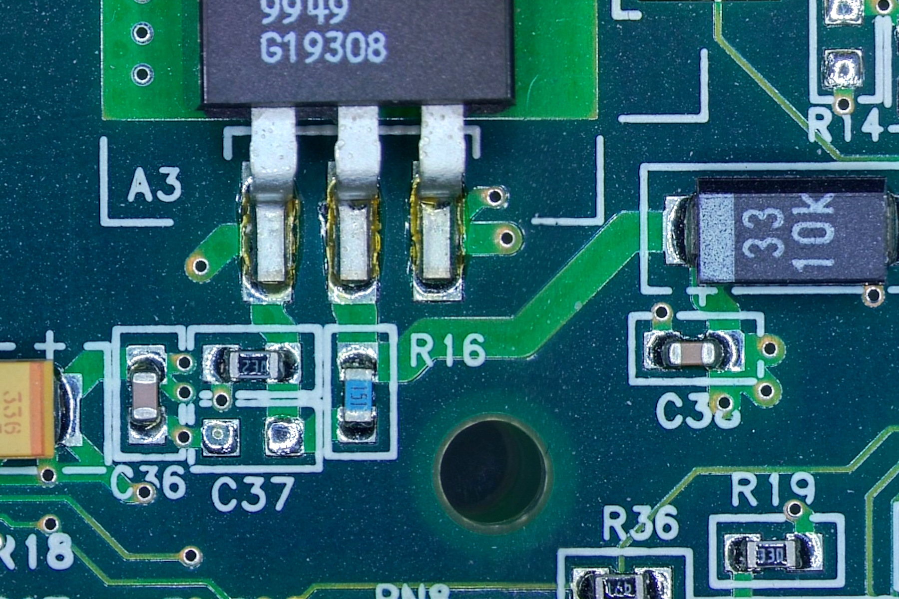
Cross-section Analysis for Electronics Manufacturing 27 Nov 2023 1:45 AM (last year)
This article describes cross-section analysis for electronics concerning quality control and failure analysis of printed circuit boards (PCBs) and assemblies (PCBAs), integrated circuits (ICs), etc.
How to Get Deeper Insights into your Organoid and Spheroid Models 22 Nov 2023 6:09 AM (last year)
In this eBook, learn about key considerations for imaging 3D cultures, such as organoids and spheroids, and discover microscopy solutions to shed new insights into dynamic processes in 3D real-time
Studying Virus Replication with Fluorescence Microscopy 15 Nov 2023 2:48 AM (last year)

The results from research on SARS-CoV-2 virus replication kinetics, adaption capabilities, and cytopathology in Vero E6 cells, done with the help of fluorescence microscopy, are described in this article.
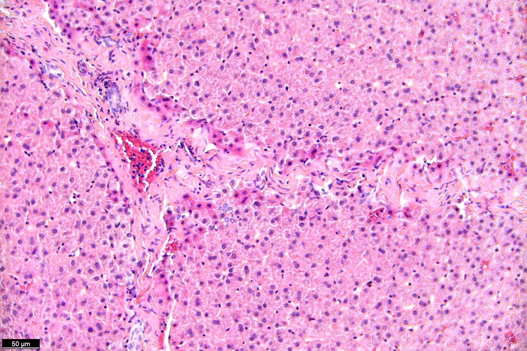
In Situ Identification of Cancer Stem Cell Niches in Hepatocellular Carcinoma 8 Nov 2023 10:10 PM (last year)
Discover how multiplexed imaging technology uncovers cancer stem cell niches in Hepatocellular Carcinoma using multiplex immunodetection, revealing extracellular matrix dynamics. Explore precise tissue insights with Cell DIVE.
Understanding Clearly the Magnification of Microscopy 7 Nov 2023 11:00 PM (last year)

To help users better understand the magnification of microscopy and how to determine the useful range of magnification values for digital microscopes, this article provides helpful guidelines.
Epi-Illumination Fluorescence and Reflection-Contrast Microscopy 2 Nov 2023 7:35 AM (last year)

This article discusses the development of epi-illumination and reflection contrast for fluorescence microscopy concerning life-science applications. Much was done by the Ploem research group collaborating with the company Leitz.
Discover how Multiplexed Bioimaging can Advance Cancer Research 1 Nov 2023 9:47 PM (last year)

Explore multiplexing with up to 60 biomarkers, enabling advanced tumor imaging approaches to gather precise, spatially-resolved single-cell data that helps enhance cancer research and clinical practice.
Posterior Segment Surgery: Benefits of Utilizing Intraoperative OCT 30 Oct 2023 11:03 PM (last year)

Learn about the value of intraoperative optical coherence tomography in posterior segment surgery to precisely locate, evaluate and manage pathologies.
Five Inverted-Microscope Advantages for Industrial Applications 24 Oct 2023 1:06 AM (last year)

With inverted microscopes, you look at samples from below since their optics are placed under the sample, with upright microscopes you look at samples from above. Traditionally, inverted microscopes are used for life science research, because gravity makes samples sink to the bottom of a holder with aqueous solution and you don’t see a lot from above.
Potential of Multiplex Confocal Imaging for Cancer Research and Immunology 23 Oct 2023 2:36 AM (last year)
Explore the new frontiers of multi-color fluorescent imaging: from image acquisition to analysis
Intraoperative OCT-Assisted Corneal Transplant Procedures 16 Oct 2023 1:01 AM (last year)

Learn about the use of intraoperative optical coherence tomography in corneal transplantation and how it facilitates the adaptation of the donor cornea.
Enhancing Neurosurgery Teaching 12 Oct 2023 12:37 AM (last year)

Learn about the Serious Game in Intraoperative Neurosurgery and how it supports neurosurgical teaching and the acquisition of decision-making skills.